















Product information
ROX Prothrombin
Transferred from the original PDF and compared with it. The original document remains authoritative.
For In Vitro Research Use – Not For Diagnostic Use
ENGLISH – Insert Revision 01/2013
INTENDED USE
For quantitative determination of Prothrombin (FII) functional activity in plasma and FII containing concentrates. The method is suitable for plasma collected in citrate or EDTA.
BIOCHEMISTRY
Factor II is a single chain vitamin K dependent glycoprotein of 72 kDa, which is activated to thrombin (FIIa) by FXa in the presence of FVa, calcium ions and phospholipids.
MEASUREMENT PRINCIPLE
FII functional activity is determined in a chromogenic prothrombinase method, in which human FII is activated to thrombin (FIIa) by human FXa in the presence of bovine FV, calcium ions and phospholipid. The amount of FIIa formed is determined from the hydrolysis of a chromogenic FIIa substrate. The FII activity of the sample is assigned vs. plasma or a FII concentrate standard with FII potency expressed in International Units (IU). The prothrombinase complex is sensitive to γ-carboxylation and therefore a-carboxy-FII is not activated in this method in contrast to snake venom based prothrombin methods.
KIT COMPOSITION
Activator Reagent, 3.0 mL (4 vials) – REF 2010
The Activator Reagent contains lyophilized human FXa, bovine FVa, CaCl₂ and phospholipids. Each vial is sufficient for 25 tests.
FIIa Substrate, 6 mL (1 vial) – REF 2080
Liquid solution of a chromogenic FIIa substrate (H-D-Phe-Pip-Arg- pNA), 2.0 mmol/L.
FII Diluent Buffer, Stock Solution, 20 mL (1 vial) – REF 2050
Liquid stock solution of diluent buffer.
PRECAUTIONS AND WARNINGS
CAUTION: Each donor unit used in the preparation of the Activator Reagent has been tested by FDA approved methods for the presence of Hepatitis B surface antigen and antibodies to HIV 1 and 2 and Hepatitis C and found to be negative. However, since no test can completely rule out the presence of these blood borne diseases, the handling and disposal of this human source reagent should be made with care.
- Avoid contact with skin and eye.
- Do not empty into drains.
- Wear suitable protective clothing.
PREPARATION
Activator Reagent
Reconstitute with 3.0 mL water. Allow to stand for 5 min at 20-25°C with intermittent gentle mixing for complete reconstitution.
FIIa Substrate, 6 mL
Ready for use.
FII Diluent Buffer, Stock Solution, 20 mL
Dilute 1 + 9 with water to obtain a 0.05 mol/L Tris-HCl buffer working solution, pH 7.3 (20°C), with 1% bovine serum albumin.
Note: All reconstitutions and dilutions should be made with water of a quality of at least NCCLS Type II water³ or Ph Eur water for injection.
STORAGE AND STABILITY
The sealed reagents are stable at 2-8°C until the Expiry Date printed on the label. Be careful to avoid contamination of the reagents by microorganisms.
- Reconstituted Activator Reagent: Stability after reconstitution is 8 hours at 2-8°C, 2 hours at 20 - 25°C and 2 hours at 37°C.
- Chromogenic FIIa substrate: Opened vial is stable for 1 month at 2-8°C.
- FII Diluent Buffer Stock Solution: Opened vial is stable for 1 month at 2-8°C. Buffer working solution should be used the same day as prepared.
MATERIALS REQUIRED BUT NOT PROVIDED
- Deionized water, NCCLS Type II water or Ph Eur water for injection or higher quality.
- Human Plasma or FII concentrate, potency assigned vs. a WHO International Standard for FII activity.
- Citric acid, 2% (for end-point method)
- Calibrated pipettes
- Photometer, 405 nm (and 490 nm for end-point method)
- Heat incubator 37°C
- Plastic test tubes and Vortex mixer
- Stop-watch
SYMBOLS USED
| Symbol | Meaning |
|---|---|
| REF | Catalog number |
| LOT | Batch code |
| Hourglass | Use by |
| Thermometer | Temperature limitation |
| Book with “i” | Consult instruction for use |
| Biohazard | Biological risks |
| Factory | Manufacturer |
METHOD - PLASMA
A calibration curve should be included in each run. A normal human plasma calibrated against a WHO International Standard should be used as calibrator. Prepare all dilutions in plastic tubes.
10.1 Preparation of standard dilutions - Plasma
Prepare independent predilutions for each standard dilution.
| Factor II % | Total Dilution | Volume of Plasma | Volume of Diluent Buffer working solution |
|---|---|---|---|
| Predilution | 1:50 | 20 µL | 980 µL |
| Factor II % | Total Dilution | Volume of Predilution | Volume of Diluent Buffer working solution |
|---|---|---|---|
| 150% | 1:133 | 300 µL of predilution | 500 µL |
| 125% | 1:160 | 200 µL of predilution | 440 µL |
| 100% | 1:200 | 100 µL of predilution | 300 µL |
| 67% | 1:300 | 100 µL of predilution | 500 µL |
| 33% | 1:600 | 50 µL of predilution | 550 µL |
| 10% | 1:2000 | 25 µL of predilution | 975 µL |
| 0% | - | - | 500 µL |
NOTE: 100% activity is defined as a FII activity of 1 IU/mL in plasma. In case the FII activity of the plasma standard differs from this value, an appropriate correction factor should be used when calculating the sample result. It is recommended to express all sample results as IU/mL.
10.2 Sample dilution - Plasma
Plasma samples should be analyzed with a sample dilution of 1:200. The FII activity of the tested sample is derived from the calibration curve.
10.3 Sample blank
Due to the high sample dilution, a sample blank does not have to be included when analyzing plasma samples.
METHOD – CONCENTRATES
A calibration curve should be included in each run. A Factor II concentrate calibrated against a WHO International Standard should be used as calibrator. Prepare all dilutions in plastic tubes.
11.1 Preparation of standard dilutions - FII containing concentrates
Prepare independent predilutions for each standard dilution.
Example, using the 3ʳᵈ International Standard for Factors II and X Concentrates 98/590 with an assigned Factor II activity of 11.2 IU/ampoule: Reconstitute one ampoule with 1.0 mL of distilled water followed by a predilution of 1:560 in Diluent Buffer-working solution to arrive at 20 mIU/mL. Prepare standard dilutions using the predilution of 20 mIU/mL according to the table below.
NOTE: The table below for preparation of standard dilutions is provided as an example only. Any dilution scheme resulting in final standard dilutions in the range 0.5 – 7.5 mIU/mL could be used.
Predilution: Predilute to 20 mIU/mL in Diluent Buffer, working solution
| Factor II mIU/mL | Volume of Predilution | Volume of Diluent Buffer working solution |
|---|---|---|
| 7.5 mIU/mL | 300 µL of predilution | 500 µL |
| 6.25 mIU/mL | 200 µL of predilution | 440 µL |
| 5 mIU/mL | 100 µL of predilution | 300 µL |
| 3.33 mIU/mL | 100 µL of predilution | 500 µL |
| 1.67 mIU/mL | 50 µL of predilution | 550 µL |
| 0.5 mIU/mL | 25 µL of predilution | 975 µL |
| 0 mIU/mL | - | 500 µL |
11.2 Sample dilution - FII containing concentrates
Prepare sample dilutions in FII Diluent Buffer working solution to obtain activities in the range 0.5 – 7.5 mIU/mL. It is recommended to analyse FII concentrate samples at several different dilutions, starting at a FII activity of about 7.5 mIU/mL, to establish the minimal dilution required to avoid any matrix interference. All dilutions should be prepared in plastic tubes.
ASSAY – PLASMA AND CONCENTRATES
The same assay procedure should be used for both plasma and concentrates.
| Step | Volume |
|---|---|
| Sample / Standard dilution (20-25°C) | 50 µL |
| Incubation 2-4 min, 37°C | |
| Activator Reagent (37°C) | 100 µL |
| Activation 15 min, 37°C | |
| FIIa Substrate (37°C) | 50 µL |
| Kinetic method: Read ΔA405/min at 37°C End-point method: Incubate at 37°C for 10 min | |
| Citric Acid, 2% (End-point method only) | 50 µL |
Kinetic reading:
Read the absorbance change at 405 nm.
End-point method:
Stop the reaction with 2% citric acid after 10 min hydrolysis at 37°C. Read the absorbance at 405 nm, using 490 nm as reference wavelength. Absorbance readings should be made within 2 hours after termination of the substrate hydrolysis.
CALCULATION
Plasmas:
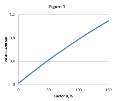
- Plot the maximal absorbance change/minute (ΔA405ₘₐₓ/min) or absorbance (A405-490) vs. FII activity in a Lin-Lin graph. Use a quadratic curve fit (Fig 1).
- The FII activity of the tested sample is obtained directly from the calibration curve. Correct the obtained value with an appropriate correction factor if the FII activity of the normal plasma standard differs from 1 IU/mL.
- If several sample dilutions are used, adjust for the dilution with the appropriate dilution factor.
- Express the sample result as IU/mL or %.
Concentrates:
The European Pharmacopoeia recommends slope ratio or parallel line evaluation for assigning activities of factor concentrates¹. Alternatively, the Factor II activity in each dilution of the tested sample can be directly obtained from the calibration curve. The result should then be multiplied by the dilution used.
- Plot the maximal absorbance change/minute (ΔA405ₘₐₓ/min) or absorbance (A405-490) vs. FII activity in a Lin-Lin graph (slope ratio evaluation) or in a Log-Log graph after subtracting the reagent blank (parallel line evaluation).
- Determine the FII activity of the sample from the calibration curve using the slope ratio or parallel line model.
- Express the sample results as IU/mL and adjust for the dilution used.
PERFORMANCE CHARACTERISTICS
The assay allows detection of about 0.25 mIU/mL (5%) FII activity.
EXPECTED VALUES
Normal Factor II levels in plasma range from 0.75 - 1.3 IU/mL.
INTERFERENCE
Factor II results are not affected at plasma levels up to the stated levels below:
| Substance | Level |
|---|---|
| Hemoglobin: | 10 mg/mL |
| Bilirubin: | 800 µg/mL |
| Triglycerides: | 10 mg/mL |
| Heparin, LMW: | 4 U/mL |
| Heparin, UF: | 4 U/mL |
REFERENCES
- 6ᵗʰ Edition of the European Pharmacopoeia, General Chapter 5.3 Statistical analysis of results of biological assays and tests.
- Kirchhof BRJ, Vermeer C, Hemker HC. The determination of prothrombin using synthetic chromogenic substrates; choice of a suitable activator. Thromb Res 13, 219-232 (1978).
- National Committee for Clinical Laboratory Standards. Specification for reagent water used in the clinical laboratory, NCCLS Approved Standard: ASC-3.
Manufacturer
Rossix AB Taljegårdsgatan 3B SE-431 53 Mölndal, Sweden info@rossix.com www.rossix.com