















Produktinformationen
MAIPA
Aus dem Original-PDF übertragen und mit ihm abgeglichen. Maßgeblich bleibt das Originaldokument.
MAIPA Assay 96T IVD REF 900006
Manufacturer: Advanced Practical Diagnostics BV, Raadsherenstraat 3, 2300 Turnhout, Belgium e-mail: admin@apdia.be web site: www.apdiagroup.com
CE 0123
INTENDED PURPOSE
The MAIPA Assay is a qualitative in vitro diagnostic medical device intended for professional laboratory use for the detection and identification of anti-platelet antibodies in serum and plasma samples and for detecting autoantibodies bound to the platelets of the patient. The test is an aid in the diagnosis of patients with identified or suspected immunological platelet disorders like Fetal-Neonatal Allo-Immune Thrombocytopenia (FNAIT), Post-Transfusion Purpura (PTP), Platelet Refractoriness (PR) and Immune Thrombocytopenias. The MAIPA Assay is designed for manual use.
DEFINITION AND SCOPE
The MAIPA Assay is based on the MAIPA (Monoclonal Antibody-specific Immobilization of Platelet Antigen) technique. This test, described by Kiefel et al (Blood 70: 1722-1726, 1987) can detect with high sensitivity and specificity anti-platelet antibodies and allows identification of the specificity of the antibody.
The assay can be used for screening serum or plasma for anti-platelet antibodies (Indirect MAIPA or MAIPAI) and/or testing for antibodies bound to the patient's platelets (Direct MAIPA or MAIPAD). Antibodies present in the serum or plasma may be alloantibodies and/or circulating autoantibodies.
A positive test result in the indirect MAIPA may be followed by the subsequent identification of the antibodies to implicated HPA-antigen using the same method.
The identification test applies for:
- patient samples tested positive for anti-platelet antibodies.
- samples of pregnant women whatever the outcome of the antibody screening test.
- samples of patients where clinical diagnosis points to platelet immunization.
The MAIPA technique is also used for cross-match reactions of:
- donor platelet antigens and serum of the recipient (PR).
- paternal platelet antigens (and/or antigens of the baby) and serum of the mother (FNAIT).
PRINCIPLE OF THE METHOD OF DETECTION AND IDENTIFICATION
The test principle is based on the capture of a platelet antigen using a mouse monoclonal antibody that reacts specifically with a single human platelet membrane glycoprotein. This is followed by binding of human antibodies to antigens on this glycoprotein and analysis of bound human IgG by an ELISA immuno-assay.
The technique is performed in two steps:
- Screening of the glycoprotein complex implicated in immunization (MAIPAI and MAIPAD)
- Identification using genotyped platelets (MAIPAI only)
For screening, platelets from a pool comprising 6 to 12 erythrocyte group O donors selected for their particular platelet genotype are incubated with the serum to be tested (indirect MAIPA) and mouse monoclonal antibodies specific for each of the four relevant platelet glycoproteins: GPIIbIIIa, GPIaIIa, GPIbIX and β-2-microglobulin/HLA. For a direct MAIPA, platelets of patients which already have an IgG bound, are directly incubated with the same set of monoclonal antibodies. Currently, 4 murine monoclonal antibodies, anti-GPIIbIIIa, anti-GPIaIIa, anti-GPIbIX and anti-β-2-microglobuline/HLA are tested in parallel in 4 individual reactions.
After incubation platelets are lysed. The lysates are then cleared by centrifugation and placed in the wells of a microplate pre-coated with goat anti-mouse IgG antibodies. The mouse monoclonal antibody – platelet glycoprotein complex specific to the potential anti-human platelet antibody is thereby attached to the bottom of the plate.
The binding of this complex is detected in a second step by a goat peroxidase-coupled anti-human IgG and revealed by the peroxidase substrate TMB (3,3’,5,5’-Tetramethylbenzidine). A blue color appears indicating the presence of an anti-GP antibody (corresponding to the specificity of the monoclonal antibody present for this reaction). The reaction is stopped by adding H₂SO₄ and the blue colour is converted into an equivalent amount of yellow colour measurable at 450 nm.
The second step consists of identifying the antibody detected in the screening test and follows the same technical protocol. Individual platelets of known genotype are selected and serum is tested with the mouse monoclonal antibody that gave a positive result in the first screening assay stage. This second step is generally performed only for the indirect test. If the direct test shows platelet bound antibodies to one or more glycoproteins, further examination to find the specific platelet antigen (HPA) is performed only in exceptional cases but not routinely.
MAIPA PROCEDURE
1. Reagents Complete MAIPA Assay kit (REF 900006)
Platelet Antibody Screening Cells
| Component | Description | Quantity, Volume | Volume needed for one test | Format |
|---|---|---|---|---|
| Screening cells SCREEN PLTL | Platelet pool of 6 to12 blood group O donors, typed for HPA-1, -2, -3, -4, -5, -6, -15 | 5 identical vials 5 x 1 ml | 50 µl | RTU |
Platelet Antibody Identification Panel Cells
| Component | Description | Quantity, Volume | Volume needed for one test | Format |
|---|---|---|---|---|
| Identification platelet 1 IDENT PLTL 1 | Single donor (blood group O) platelet cells, typed for HPA-1, -2, -3, -4, -5, -6, -15; batch dependent | 1 vial, 0,65 ml | 50 µl | RTU |
| Identification platelet 2 IDENT PLTL 2 | Single donor (blood group O) platelet cells, typed for HPA-1, -2, -3, -4, -5, -6, -15; batch dependent | 1 vial, 0,65 ml | 50 µl | RTU |
| Identification platelet 3 IDENT PLTL 3 | Single donor (blood group O) platelet cells, typed for HPA-1, -2, -3, -4, -5, -6, -15; batch dependent | 1 vial, 0,65 ml | 50 µl | RTU |
| Identification platelet 4 IDENT PLTL 4 | Single donor (blood group O) platelet cells, typed for HPA-1, -2, -3, -4, -5, -6, -15; batch dependent | 1 vial, 0,65 ml | 50 µl | RTU |
| Identification platelet 5 IDENT PLTL 5 | Single donor (blood group O) platelet cells, typed for HPA-1, -2, -3, -4, -5, -6, -15; batch dependent | 1 vial, 0,65 ml | 50 µl | RTU |
| Identification platelet 6 IDENT PLTL 6 | Single donor (blood group O) platelet cells, typed for HPA-1, -2, -3, -4, -5, -6, -15; batch dependent | 1 vial, 0,65 ml | 50 µl | RTU |
Kit Controls
| Component | Description | Quantity, Volume | Volume needed for one test | Format |
|---|---|---|---|---|
| Control serum/plasma CONTR 1a | Anti-HPA-1a positive | 1 vial, 0,4 ml | 50 µl | RTU |
| Control serum/plasma CONTR 5b | Anti-HPA-5b positive | 1 vial, 0,4 ml | 50 µl | RTU |
| Control serum/plasma CONTR HLA | Anti-HLA positive | 1 vial, 0,4 ml | 50 µl | RTU |
| Control serum/plasma CONTR NEG | Negative | 3 vials, 3 x 0,4 ml | 50 µl | RTU |
MAIPA Reagents
| Component | Description | Quantity, Volume | Volume needed for one test | Format |
|---|---|---|---|---|
| Microplate MTP | 12 individual strips | 1 plate, 96 wells | RTU | |
| Antibody MAB IIbIIIa | Anti-GPIIbIIIa | 2 vials, 1,2 ml | 50 µl | RTU |
| Antibody MAB IaIIa | Anti-GPIaIIa | 1 vial, 1,8 ml | 50 µl | RTU |
| Antibody MAB IbIX | Anti-GPIbIX | 1 vial, 1,5 ml | 50 µl | RTU |
| Antibody MAB HLA | Anti-HLA (B2M) | 1 vial, 1,5 ml | 50 µl | RTU |
| Cell Wash Buffer CELLWASHBUF 10x | MAIPA Platelet Wash Buffer | 1 vial, 20 ml | (2+2+4) x 200 µl | 10x |
| Platelet Lysis buffer LYSBUF | MAIPA Platelet Lysis Buffer | 1 vial, 15 ml | 130 µl | RTU |
MAIPA ELISA Reagents
| Component | Description | Quantity, Volume | Volume needed for one test | Format |
|---|---|---|---|---|
| Coated microtiterplate COATMTP | Goat anti-mouse IgG coated microtiter plate | 1 plate, 96 wells | RTU | |
| Conjugate CONJ | Goat anti-Human IgG-HRP | 1 vial, 12 ml | 100 µl | RTU |
| ELISA Wash buffer ELISAWASHBUF 20x | TRIS buffered Triton X-100 / Tween 20 | 1 vial, 50 ml | 12 x 200 µl | 20x |
| Chromogen TMB TMB | TMB | 1 vial, 12 ml | 100 µl | RTU |
| Stop Solution STOPSOL | 0.5 M H₂SO₄ | 1 vial, 12 ml | 100 µl | RTU |
2. Additional reagents required but not provided by the manufacturer
- Distilled water (for buffer dilution)
- PBS 1x w/1 % BSA, w/ 0,33 % EDTA (for platelets preparation in Direct MAIPA)
3. Further materials required but not provided by the manufacturer
- Micropipettes 10-100 µl and tips
- Multi-channel pipette100-250 µl and tips
- Tubes and vials for reagents dilution
- Reagent reservoirs
- Vortex mixer
- Microplate shaker
- Microplate reader for measurement of optical densities at 450 nm with reference filter 600-650 nm
- Microplate centrifuge validated for MAIPA (see section 8)
- Incubator at 36 ± 1 °C
- Refrigerator at 2 – 8 °C
- Absorbent paper
4. Warnings and precautions for users
4.1. For in vitro diagnostic use only.
4.2. For professional laboratory use.
4.3. Do not mix reagents or coated microtiter strips from kits with different lot numbers.
4.4. Treat controls and samples as if they contain infectious agents.
4.5. Dispose of patient samples and all materials used to perform the test as if contaminated with potentially infectious substances. Safe disposal must be in accordance with hospital policies and local and/or national legislation.
4.6. Stop Solution is a 0,5 M H2SO4 solution which is irritant. In case of contact with eyes or skin, rinse with plenty of water and seek medical advice.
4.7. The Lysis Buffer and the ELISA Wash Buffer 20x contain the substance Triton X-100. Triton X-100 is a surfactant belonging to the group of OPEs (octylphenol ethoxylates) that have been included in Annex XIV (Authorisation list) of REACH Regulation (EC) N° 1907/2006. These substances have endocrine disrupting properties through their degradation, for which it is scientifically proven that they can have serious effects on the environment.
Classification for Lysis Buffer :
Signal word : Warning
Hazard pictograms:

Following hazard statements are applicable : H317 : May cause an allergic skin reaction. H411 : Toxic to aquatic life with long lasting effects.
Following precautionary statements are applicable : P273 : Avoid release to the environment. P280 : Wear protective gloves/protective clothing/eye protection/face protection/hearing protection/… P302+P352 : IF ON SKIN : Wash with plenty of water/… P391 : Collect spillage.
Classification for ELISA Wash Buffer 20x:
Signal word : Danger
Hazard pictograms:

Following hazard statements are applicable : H317 : May cause an allergic skin reaction. H318 : Causes serious eye damage. H315 : Causes skin irritation. H410 : Very toxic to aquatic life with long lasting effects.
Following precautionary statements are applicable : P273 : Avoid release to the environment. P280 : Wear protective gloves/protective clothing/eye protection/face protection/hearing protection/… P302+P352 : IF ON SKIN : Wash with plenty of water/… P310 : Immediately call a POISON CENTER/doctor/… P391 : Collect spillage.
4.8. Chromogen Solution contains the hazardous ingredient N-Methyl-2-pyrrolidone at a concentration > 0,3 %. It is classified as a Reproductive Toxicant Category 1B. Signal word: Danger
Hazard pictogram:

Following hazard statements are applicable: H360D: May damage the unborn child.
Following precautionary statements are applicable: P280: Wear protective gloves/protective clothing/eye protection/face protection/hearing protection/… P308+P313: IF exposed or concerned: Get medical advice/attention.
4.9. Some kit components contain sodium azide as a preservative. In order to prevent the formation of potentially explosive metal azides in laboratory plumbing, flush drains thoroughly after disposal of these solutions.
4.10. After obtaining the results, no decisions should be made without first consulting the medical relevance of these results.
4.11. Serious incidents related to the MAIPA Assay must be reported to the manufacturer.
5. Sample material
Serum or plasma can be used for indirect MAIPA. Specimens can be stored at 2 – 8 °C for 3 to 4 days or they can be stored frozen at – 20 °C for at least one year. Frozen samples can be subsequently thawed and refrozen three times (three freeze/thaw cycles) without any impact on the results with regard of correct detection of anti-platelet antibodies.
Platelets isolated from EDTA whole blood can be used for direct MAIPA. Whole blood is centrifuged at 200g for 10 minutes to form platelet-rich plasma (PRP). The PRP is then centrifuged at 2050g for 10 minutes to pellet the platelets. After removal of the plasma, the cells are washed twice with PBS / 1% BSA / 0.33% EDTA and adjusted to 500.10⁶ cells/ml in the same buffer (the recommended minimum amount of platelets for one autologous test is 5.10⁶). The prepared patient platelet suspension can be stored at 2 – 8 °C for maximum 5 days.
6. Controls
Negative and positive control samples should be included in each determination to validate the results. Two blank reagent control wells should be included in all test procedures.
A set of 4 control Plasma/Sera (CONTR 1a, CONTR 5b, CONTR HLA, CONTR NEG) is included in the MAIPA Assay kit.
7. Reagent preparation and storage conditions
This device is made for single use but the reagents supplied with the kit can be used during multiple runs using part of the provided reagents in each run. All reagents must be kept at 2 – 8 °C. All reagents can be stored at 2 – 8 °C until the expiry date indicated on the label. Never use any kit components beyond the indicated expiry date.
The complete MAIPA procedure takes about 6 hours: the required amount of all reagents will be removed from the refrigerator immediately before use. An environment temperature in the lab of 19-25°C is advised. The remaining unused reagents should be re-stored at 2-8°C as soon as possible. Reagents should be used within six months after the first opening.
Both 10x and 20x concentrated wash buffers have to be diluted to 1x strength before starting the assay. Diluted wash buffers can be kept at room temperature during the time of the test. Store remaining volume of the 1x diluted buffers at 2 – 8 °C after the test. They can be reused for maximum 1 month when intermittently stored at 2 – 8 °C.
10x concentrated Cell Wash Buffer can contain phosphate crystals after storage at 2 - 8 °C. These crystals will disappear at room temperature. Wait for diluting the Cell Wash buffer until no crystals remain.
8. Test procedure: general information
MICROPLATE METHOD for platelet incubation and washings!
Samples may be tested in singlicate or in duplicate.
A microplate washer may be used but in this case the washing steps should be validated. If necessary increase the number of washing cycles.
All centrifugation steps are done at room temperature: 19-25°C. It is important to validate your centrifuge for usability with regard of the MAIPA procedure.
A centrifuge equipped with a swing-out rotor and four microplate holders positioned in the rotor cross at an angle of 45 °C to the rotor arms is preferred. Example: Eppendorf 5810 with rotor A-4-81and MTP buckets or with rotor A-4-62 with MTP buckets. We do not recommend a centrifuge equipped with a swing-out rotor and two microplate holders positioned in parallel with the rotor arm. Example: Eppendorf 5430 with rotor A-2-MTP.
Beside the use of either screening or identification cells, the assay procedure is identical for both screening and identification of anti-platelet antibodies. As such screening and identification can be performed simultaneously on the same plate.
The entire MAIPA procedure is given below.
9. Test procedure Indirect MAIPA (MAIPAI): detailed description
9.1. Reagent preparation
9.1.1. Dilute (1/10) CELL Wash Buffer (CELLWASHBUF 10x) in distilled water and keep it at room temperature (19-25°C).
9.1.2. Dilute (1/20) ELISA Wash Buffer (ELISAWASHBUF 20x) in distilled water and keep it at room temperature (19-25°C).
9.2. Preparation of the platelets
FOR SCREENING OF ANTI-PLATELET ANTIBODIES
9.2.1. s Remove the uncoated MICROPLATE (MTP) from the box. Push the 8 micro-well rows out of the frame according to the necessary number of wells (don’t forget the controls). Place these micro-wells into an empty frame. Put the remaining micro-wells/plate back in the box. Provide as many wells as patients and targeted glycoproteins to be tested. Generally 8 wells are required per patient for an anti-platelet antibody screening test, which consists of both a direct and an indirect MAIPA. For both of these assays 4 glycoproteins (GPs) are tested in 4 separate wells. For each screening assay, provide 6 extra wells for the controls in order to validate the run: 2 blank reagent wells on position A1 and B1; 3 positive controls plasma/serum in wells C1 (anti-HPA-1a), D1 (anti-HPA-5b) and E1 (anti-HLA); and 1 negative control plasma/serum in well F1.
9.2.2. s Add 50 µl of SCREENING CELLS (SCREEN PLTL) in the sample and control wells. The Platelet Antibody Screening Cells are ready-to-use.
9.2.3. s Centrifuge the MICROPLATE (MTP) at 1050g ± 50g for 3 minutes. Empty the wells by inverting the microplate taking care to keep the cells at the bottom of the wells. Mix the platelets gently using a microplate shaker (2 x 10 seconds at 700-1000 rpm; first 10 seconds in one direction, followed by 10 seconds in the opposite direction).
9.2.4. s Wash the MICROPLATE (MTP) as follows: add 200 µl of diluted CELL WASH BUFFER in each well and centrifuge at 1050g ± 50g for 3 minutes. Empty the wells by inverting the microplate taking care to keep the cells at the bottom of the wells. Mix the platelets gently using a microplate shaker (2 x 10 seconds at 700-1000 rpm as described previously).
9.2.5. s Repeat step 9.2.4. s once (total of 2 washes).
FOR IDENTIFICATION OF ANTI-PLATELET ANTIBODIES
9.2.1. i Remove the uncoated MICROPLATE (MTP) from the box and proceed as described in 9.2.1.s. For an anti-platelet antibody identification MAIPA (always indirect) 6 wells are required per positive test result obtained in the preceding screening assay. One sample may react with several monoclonal antibodies. Generally, identification is done with anti-GPIaIIa and/or GPIIbIIIa. Identification is not performed for samples that reacted positive for anti-HLA antibodies in the screening assay. If the identification is performed in a separate assay, provide 6 extra wells for the controls in each run for validation: 2 blank reagent wells on position A1 and B1; 3 positive control plasma/sera in wells C1 (anti-HPA-1a), D1 (anti-HPA-5b) and E1 (anti-HLA); and 1 negative control plasma/serum in well F1. Low volume laboratories might prefer to limit the number of controls used. In this case, we would advise to use only 3 control wells: 1 blank reagent well on position A1; 1 positive control in well B1; and 1 negative control in well C1. For the positive control it is suggested to use the control that reacts with the monoclonal antibody giving rise to the positive test result in screening step.
9.2.2. i For an anti-platelet antibody identification (indirect) MAIPA add 50 µl of each of the six IDENTIFICATION PLATELETS (IDENT PLTL) in the respective sample wells. Add 50 µl of SCREENING CELLS (SCREEN PLTL) in the control wells. The Platelet Antibody Screening Cells and Platelet Antibody Identification Panel Cells are ready-to-use.
9.2.3. i Proceed with washing of the IDENTIFICATION PLATELETS (IDENT PLTL) according to 9.2.3.s till 9.2.5.s.
9.3. Incubation of platelets with serum
FOR SCREENING OF ANTI-PLATELET ANTIBODIES
9.3.1. s For an anti-platelet antibody screening assay (indirect MAIPA) add 50 µl of patient plasma/serum in 4 wells containing SCREENING CELLS (SCREEN PLTL). Add 50 µl of the CONTROL PLASMA/SERA (CONTR) in the respective wells.
9.3.2. s Mix gently manually or by using a microplate shaker (2 x 10 seconds at 700-1000 rpm as described previously).
9.3.3. s Incubate for 30 ± 5 minutes at 36 ± 1 °C.
9.3.4. s Centrifuge the MICROPLATE (MTP) at 1050g ± 50g for 3 minutes. Empty the wells by inverting the microplate taking care to keep the cells at the bottom of the wells. Mix the platelets gently using a microplate shaker (2 x 10 seconds at 700-1000 rpm as described previously).
FOR IDENTIFICATION OF ANTI-PLATELET ANTIBODIES
9.3.1. i For an anti-platelet antibody identification assay (indirect MAIPA) add 50 µl of patient plasma/serum in 6 wells containing the different IDENTIFICATION PLATELETS (IDENT PLTL). Add 50 µl of the CONTROL PLASMA/SERA (CONTR) in the respective wells.
9.3.2. i Mix gently manually or by using a microplate shaker (2 x 10 seconds at 700-1000 rpm as described previously).
9.3.3. i Incubate for 30 ± 5 minutes at 36 ± 1 °C.
9.3.4. i Centrifuge the MICROPLATE (MTP) at 1050g ± 50g for 3 minutes. Empty the wells by inverting the microplate taking care to keep the cells at the bottom of the wells. Mix the platelets gently using a microplate shaker (2 x 10 seconds at 700-1000 rpm as described previously).
9.4. Removal of unbound immunoglobulins
9.4.1. Wash the MICROPLATE (MTP) as follows: add 200 µl of diluted CELL WASH BUFFER in each well and centrifuge at 1050g ± 50g for 3 minutes. Empty the wells by inverting the microplate taking care to keep the cells at the bottom of the wells. Mix the platelets gently using a microplate shaker (2 x 10 seconds at 700-1000 rpm as described previously).
9.4.2. Repeat step 9.4.1. once (total of 2 washes).
9.5. Incubation of platelets with monoclonal antibody
FOR SCREENING OF ANTI-PLATELET ANTIBODIES
9.5.1. s For an anti-platelet antibody screening assay add 50 µl of each MONOCLONAL ANTIBODY (MAB) in the 4 respective wells containing SCREENING CELLS (SCREEN PLTL). For controls: add 50 µl of anti-GPIIbIIIa (MAB IIbIIIa) in well C1 (CONTR 1a), 50 µl of anti-GPIaIIa (MAB IaIIa) in well D1 (CONTR 5b), 50 µl of anti-HLA/β2-microglobulin (MAB HLA) in well E1 (CONTR HLA). In well F1 (CONTR NEG), add 50 µl of anti-GPIbIX (MAB IbIX) or 50 µl of anti-GPIIbIIIa (MAB IIbIIIa) or 50 µl of anti-GPIaIIa (MAB IaIIa).
9.5.2. s Mix gently manually or by using a microplate shaker (2 x 10 seconds at 700-1000 rpm as described previously).
9.5.3. s Incubate for 30 ± 5 minutes at 36 ± 1 °C.
9.5.4. s Centrifuge the MICROPLATE (MTP) at 1050g ± 50g for 3 minutes. Empty the wells by inverting the microplate taking care to keep the cells at the bottom of the wells. Mix the platelets gently using a microplate shaker (2 x 10 seconds at 700-1000 rpm as described previously).
FOR IDENTIFICATION OF ANTI-PLATELET ANTIBODIES
9.5.1. i For an anti-platelet antibody identification assay add 50 µl of the MONOCLONAL ANTIBODY (MAB) (giving rise to a positive result in a screening test) in each of 6 wells containing IDENTIFICATION PLATELETS (IDENT PLTL). For controls: add 50 µl of anti-GPIIbIIIa (MAB IIbIIIa) in well C1 (CONTR 1a), 50 µl of anti-GPIaIIa (MAB IaIIa) in well D1 (CONTR 5b), 50 µl of anti-HLA/β2-microglobulin (MAB HLA) in well E1 (CONTR HLA). In well F1 (CONTR NEG), add 50 µl of anti-GPIbIX (MAB IbIX) or 50 µl of anti-GPIIbIIIa (MAB IIbIIIa) or 50 µl of anti-GPIaIIa (MAB IaIIa). For low volume laboratories that use a reduced number of controls (see 9.2.1): Add 50 µl of the MONOCLONAL ANTIBODY (MAB) corresponding to the chosen positive control in well B1 (e.g. anti-GPIIbIIIa for the CONTR 1a; anti-GPIaIIa for the CONTR 5b). Add 50 µl of the same MONOCLONAL ANTIBODY (MAB) in well C1 for the negative control.
9.5.2. i Mix gently manually or by using a microplate shaker (2 x 10 seconds at 700-1000 rpm as described previously).
9.5.3. i Incubate for 30 ± 5 minutes at 36 ± 1 °C.
9.5.4. i Centrifuge the MICROPLATE (MTP) at 1050g ± 50g for 3 minutes. Empty the wells by inverting the microplate taking care to keep the cells at the bottom of the wells. Mix the platelets gently using a microplate shaker (2 x 10 seconds at 700-1000 rpm as described previously).
9.6. Removal of unbound monoclonal antibody
9.6.1. Wash the MICROPLATE (MTP) as follows: add 200 µl of diluted CELL WASH BUFFER in each well and centrifuge at 1050g ± 50g for 3 minutes. Empty the wells by inverting the MICROPLATE (MTP) taking care to keep the cells at the bottom of the wells. Mix the platelets gently using a microplate shaker (2 x 10 seconds at 700-1000 rpm as described previously).
9.6.2. Repeat step 9.6.1. three times (total of 4 washes).
9.7. Solubilisation of platelet membranes
9.7.1. Add 130 µl of PLATELET LYSIS BUFFER (LYSBUF) in all wells and resuspend the platelet pellet first by gently pipetting up and down. Next, pipet the platelet suspension 10 times vigorously up and down. This handling induces foaming, which is necessary for efficient lysis of the platelets. Take good care not to cross-contaminate wells.
9.7.2. Incubate for at least 15 minutes at 2 - 8 °C.
9.7.3. Centrifuge at 1050g ± 50g for 15 minutes. Immune complexes (MAB, GP and human Ab) remain in the supernatant, while cell debris sediments to the bottom of the well.
Remark: before or after centrifugation, lysates can be stored at 2 – 8 °C °C for one night if necessary.
9.8. Transfer of the platelet lysates to the goat anti-mouse IgG coated microplate
9.8.1. Remove the goat anti-mouse IgG COATED MICROPLATE (COATMTP) from the aluminium pouch. Push the 8 micro-well rows out of the frame and cut to break off the number of wells corresponding to the number of lysates to be analysed. Place these micro-wells into an empty frame. Put the remaining micro-wells/plate back in the aluminium pouch and re-seal.
9.8.2. Transfer 100 µl of the cell lysis supernatant into the goat anti-mouse IgG coated wells without touching or aspirating the debris at the bottom of the uncoated plate.
9.8.3. Incubate for 30 ± 5 minutes at 36 ± 1 °C.
9.9. Removal of unbound lysate proteins
9.9.1. Empty the wells by inverting the MICROPLATE (COATMTP) and tap the plate on absorbent paper.
9.9.2. Wash the MICROPLATE (COATMTP) as follows: add 200 µl of diluted ELISA WASH BUFFER in each well, empty the wells by inverting the microplate and tap the plate on absorbent paper.
Remark: It is recommended to use reverse pipetting for the washing steps in order to avoid bubbling of the washing solution (risk for cross-contamination).
9.9.3. Repeat step 9.9.2. five times (total of 6 washes).
9.9.4. Make sure the wells are dry after the final wash step.
9.10. Addition of peroxidase labeled goat anti-human IgG
9.10.1. Add 100 µl of the goat anti-human IgG HRP CONJUGATE (CONJ) in each well.
9.10.2. Incubate for 30 ± 5 minutes at 36 ± 1 °C.
9.11. Removal of unbound peroxidase labeled goat anti-human IgG
9.11.1. Empty the wells by inverting the MICROPLATE (COATMTP) and tap the plate on absorbent paper.
9.11.2. Wash the microplate as follows: add 200 µl of diluted ELISA WASH BUFFER in each well, empty the wells by inverting the microplate and tap the plate on absorbent paper.
Remark: It is recommended to use reverse pipetting for the washing steps in order to avoid bubbling of the washing solution (risk for cross-contamination).
9.11.3. Repeat step 9.11.2. five times (total of 6 washes).
9.11.4. Make sure the wells are dry after the final wash step.
9.12. Addition of TMB substrate
9.12.1. Add 100 µl of the TMB solution (CHROM) in each well.
Remark: Keep the solution strictly protected from light.
9.12.2. Incubate for 15 minutes at 36 ± 1°C in the dark.
9.13. Addition of acid to stop colour development
9.13.1. Add 100 µl of STOP SOLUTION (STOPSOL) (H₂SO₄) in each well.
9.14. Reading of microtiter plate
9.14.1. Measure the optical density at 450 m with reference filter 600-650 nm in a microplate reader and record the results.
10. Test procedure Direct MAIPA (MAIPAD)
The test procedure for a direct MAIPA is generally the same as the procedure for an indirect MAIPA as described in section 9.
In the first part of the MAIPA, a prepared suspension of the patient’s platelets are used in stead of the screening platelets of the kit.
Antibodies detected during the direct MAIPA are autoantibodies directed against the glycoproteins (GPIaIIa, GPIIbIIIa or GPIbIX) on the patient’s platelets, targeting epitopes which are commonly present on the various platelet glycoproteins and not a specific HPA platelet antigen. Therefore, further identification with the identification panel cells of the MAIPA kit is not deployed.
10.1. Preparation of the platelets
10.1.1. Remove the uncoated MICROPLATE (MTP) from the box. Push the 8 micro-well rows out of the frame according to the necessary number of wells (don’t forget the controls). Place these micro-wells into an empty frame. Put the remaining micro-wells/plate back in the box. Provide as many wells as patients and targeted glycoproteins to be tested. Generally 8 wells are required per patient for an anti-platelet antibody screening test, which consists of both a direct and an indirect MAIPA. For both of these assays 4 glycoproteins (GPs) are tested in 4 separate wells. For each screening assay, provide 6 extra wells for the controls in order to validate the run: 2 blank reagent wells on position A1 and B1; 3 positive controls plasma/serum in wells C1 (anti-HPA-1a), D1 (anti-HPA-5b) and E1 (anti-HLA); and 1 negative control plasma/serum in well F1.
10.1.2. Add 50 µl of SCREENING CELLS (SCREEN PLTL) in the control wells. The Platelet Antibody Screening Cells are ready-to-use. Add 50 µl of patient platelets previously washed (2 times) and adjusted in PBS / 1% BSA / 0.33% EDTA (minimum 5 x 10⁶ platelets per test) in the sample wells. The necessary quantity of platelets to add is normally 25 x 10⁶, however, 5 x 10⁶ cells might be sufficient for thrombopenic samples. The patient platelets could also be added at the next step (when adding serum or plasma). They have been washed previously and it is not necessary to wash again.
10.1.3. Centrifuge the MICROPLATE (MTP) at 1050g ± 50g for 3 minutes. Empty the wells by inverting the microplate taking care to keep the cells at the bottom of the wells. Mix the platelets gently using a microplate shaker (2 x 10 seconds at 700-1000 rpm; first 10 seconds in one direction, followed by 10 seconds in the opposite direction).
10.1.4. Wash the MICROPLATE (MTP) as follows: add 200 µl of diluted CELL WASH BUFFER in each well and centrifuge at 1050g ± 50g for 3 minutes. Empty the wells by inverting the microplate taking care to keep the cells at the bottom of the wells. Mix the platelets gently using a microplate shaker (2 x 10 seconds at 700-1000 rpm as described previously).
10.1.5. Repeat step 10.1.4. once (total of 2 washes).
10.2. Incubation of platelets with serum
10.2.1. A direct MAIPA doesn’t require the addition of plasma/serum (antibodies are already bound to the platelets), but 50 µl of diluted CELL WASH BUFFER should be added to the sample wells to protect the cells from drying. Or patient platelets can be added at this step if it wasn’t done before (step 10.1.2.). Add 50 µl of the CONTROL PLASMA/SERA (CONTR) in the respective wells.
10.2.2. Mix gently manually or by using a microplate shaker (2 x 10 seconds at 700-1000 rpm as described previously).
10.2.3. Incubate for 30 ± 5 minutes at 36 ± 1 °C.
10.2.4. Centrifuge the MICROPLATE (MTP) at 1050g ± 50g for 3 minutes. Empty the wells by inverting the microplate taking care to keep the cells at the bottom of the wells. Mix the platelets gently using a microplate shaker (2 x 10 seconds at 700-1000 rpm as described previously).
10.3. Removal of unbound immunoglobulins
10.3.1. Wash the MICROPLATE (MTP) as follows: add 200 µl of diluted CELL WASH BUFFER in each well and centrifuge at 1050g ± 50g for 3 minutes. Empty the wells by inverting the microplate taking care to keep the cells at the bottom of the wells. Mix the platelets gently using a microplate shaker (2 x 10 seconds at 700-1000 rpm as described previously).
10.3.2. Repeat step 10.3.1. once (total of 2 washes).
10.4. Incubation of platelets with monoclonal antibody
10.4.1. For an anti-platelet antibody screening assay add 50 µl of each MONOCLONAL ANTIBODY (MAB) in the 4 respective wells containing patient platelets. For controls: add 50 µl of anti-GPIIbIIIa (MAB IIbIIIa) in well C1 (CONTR 1a), 50 µl of anti-GPIaIIa (MAB IaIIa) in well D1 (CONTR 5b), 50 µl of anti-HLA/β2-microglobulin (MAB HLA) in well E1 (CONTR HLA). In well F1 (CONTR NEG), add 50 µl of anti-GPIbIX (MAB IbIX) or 50 µl of anti-GPIIbIIIa (MAB IIbIIIa) or 50 µl of anti-GPIaIIa (MAB IaIIa).
10.4.2. Mix gently manually or by using a microplate shaker (2 x 10 seconds at 700-1000 rpm as described previously).
10.4.3. Incubate for 30 ± 5 minutes at 36 ± 1 °C.
10.4.4. Centrifuge the MICROPLATE (MTP) at 1050g ± 50g for 3 minutes. Empty the wells by inverting the microplate taking care to keep the cells at the bottom of the wells. Mix the platelets gently using a microplate shaker (2 x 10 seconds at 700-1000 rpm as described previously).
10.5. Removal of unbound monoclonal antibody
10.5.1. Wash the MICROPLATE (MTP) as follows: add 200 µl of diluted CELL WASH BUFFER in each well and centrifuge at 1050g ± 50g for 3 minutes. Empty the wells by inverting the MICROPLATE (MTP) taking care to keep the cells at the bottom of the wells. Mix the platelets gently using a microplate shaker (2 x 10 seconds at 700-1000 rpm as described previously).
10.5.2. Repeat step 10.5.1. three times (total of 4 washes).
10.6. Solubilisation of platelet membranes
10.6.1. Add 130 µl of PLATELET LYSIS BUFFER (LYSBUF) in all wells and resuspend the platelet pellet first by gently pipetting up and down. Next, pipette the platelet suspension 10 times vigorously up and down. This handling induces foaming, which is necessary for efficient lysis of the platelets. Take good care not to cross-contaminate wells.
10.6.2. Incubate for at least 15 minutes at 2 - 8 °C.
10.6.3. Centrifuge at 1050g ± 50g for 15 minutes. Immune complexes (MAB, GP and human Ab) remain in the supernatant, while cell debris sediments to the bottom of the well.
Remark: before or after centrifugation, lysates can be stored at 4 °C for one night if necessary.
10.7. ELISA part of the MAIPA procedure
Continue with the ELISA part of the MAIPA procedure as described in sections 9.8 till 9.14.
11. Concise version of the MAIPA procedure
A concise overview of the complete MAIPA procedure is added as annex to this IFU.
INTERPRETATION AND VALIDATION OF THE TEST RESULTS
Assay validation is based on the OD values of the control samples. In general it can be said that (after subtraction of the blank value):
- The OD value for the negative control should be below 0,1
- The OD value for the positive controls should be above 0,5
Interpretation of assay results is relatively straightforward with a cut-off of OD = 0,2 (after subtraction of the blank value):
- OD values above 0,2 are considered positive
- OD values below 0,2 are considered negative
It is however strongly advised for each laboratory to determine its own cut-off using a panel of negative samples, as well as the acceptance OD criteria for the negative and positive controls. Where homemade controls are used, each laboratory should define the corresponding OD values for assay validation.
- For a platelet antibody screening test, one pool of cells is used and 4 reactions with four different monoclonal antibodies are performed. If these reactions are negative no further action is required. If one of the reactions yields a positive result, a platelet antibody identification assay is performed using the monoclonal antibody that resulted in a positive screening test.
- For a platelet antibody identification assay, at least 4 cells from the platelet cell panel showing a positive platelet antibody screening phenotype and the glycoprotein-specific monoclonal antibody yielding a positive result in the preceding screening assay are used.
PERFORMANCE CHARACTERISTICS
1. Analytical Specificity
1.1. Cross-reactivity
Inherent to the localization of the glycoproteins on the membrane of the platelets, patient samples with a high titer for anti-HLA antibodies might show false positive reactions due to cross-reactivity when assayed with monoclonal detection antibody anti-GPIbIX. The nature of these false positive reactions can be examined by retesting the involved sample with chloroquine-treated screening platelets of the kit (Ref. 6). A work procedure for Chloroquine treatment is available on request.
1.2. Interference
Potential interference of endogenous substances in the MAIPA procedure has been examined for hemolytic, lipemic and icteric samples. No interference from hemoglobin (5 mg/ml), lipids (intralipid, 2,5 mg/ml) and bilirubin (0,2 mg/ml) could be found. Potential interference of exogenous substances has not been included in the study.
2. Accuracy
2.1. Trueness
Trueness of measurement has been assessed by making a series of dilutions prepared from four certified WHO Reference Reagents for detection of anti-platelet antigen antibodies (NIBSC 03/190, anti-HPA-3a; NIBSC 05/106, anti-HPA-1a; NIBSC 99/666, anti-HPA-5b and NIBSC 07/214, anti-HLA) and analyzing the dilutions in a MAIPA test to determine the dilution point for which the measured value near the cut-off value can still be considered as a true positive value. Based on these test results, the MAIPA Assay is still able to measure the WHO reference samples as a true positive value (OD > 0,200) at the by NIBSC prescribed minimum detectable dilution.
2.2. Precision
Precision of the MAIPA Assay has been investigated by assessing repeatability and reproducibility of measurements performed with the device.
Repeatability (replicate-to-replicate variability) has been assessed by analysing five samples (four positive, one negative) in 10 independent runs on ten days (four replicates per sample/run).Performed by one operator and for one batch of the MAIPA Assay. Reproducibility (combining site-to-site and operator-to-operator variability) has been assessed by analysing the same samples by a second operator in another lab on five days coinciding with days that runs for the repeatability assessment were performed.
Obtained results:
| SAMPLE | REPEATABILITY Mean (OD) | REPEATABILITY SD (OD) | REPEATABILITY CV (%) | REPRODUCIBILITY Mean (OD) | REPRODUCIBILITY SD (OD) | REPRODUCIBILITY CV (%) |
|---|---|---|---|---|---|---|
| S1 (NIBSC 03/190) | 0,698 | 0,0571 | 8,2 | 0,675 | 0,1327 | 19,7 |
| S2 (NIBSC 05/106) | 0,441 | 0,0378 | 8,6 | 0,414 | 0,0561 | 13,6 |
| S3 (NIBSC 99/666) | 1,029 | 0,0951 | 9,2 | 1,029 | 0,1214 | 11,8 |
| S4 (NIBSC 03/190 + NIBSC 07/214, 1:1) | 1,061 | 0,1114 | 10,5 | 0,970 | 0,1037 | 10,7 |
| S5 (CONTR NEG) | 0,065 | 0,0101 | 15,5 | 0,068 | 0,0113 | 16,7 |
3. Cut-off
A sample panel consisting of 30 samples positive for a variety of platelet antibodies and 30 samples negative for the presence of platelet antibodies was analysed in the MAIPA Assay. Based on the known clinical state of the samples and the measured ODs in the test, a ROC Curve was constructed. Applying the cut-off value OD < 0.200, sensitivity and specificity were both calculated as 96,67 % (95 % CI: 83.33 to 99,41 %).
4. Clinical performance
In a validation study in a French Reference Lab for Platelet Immunology, 30 archived patient samples (15 sera and 15 plasma samples from patients from the target population, diagnosed with FNAIT, PTP and PR) previously tested positive for anti-platelet antibodies by MAIPA technology, were analysed again using the MAIPA Assay. One previously weak positive sample was found negative in the test. A diagnostic sensitivity of 96.7 % was obtained.
In the same study, 210 negative samples from 105 donors (matched serum and plasma specimens) with no anti-platelet antibodies were analysed in the MAIPA Assay. A diagnostic specificity of 98,5 % was obtained.
In a comparative study, a sample panel was analysed with the MAIPA Assay and the Pak Lx™ Assay (Immucor GTI). The sample panel consisted of 30 samples positive for anti-platelet antibodies and 30 negative samples. Due to bead failure in the Pak Lx™ Assay, four test results had to be omitted.
Following results were obtained for the remaining 56 samples (28 positive, 28 negative):
| Pak Lx (Candidate Method) | MAIPA (Reference Method): Positive | MAIPA (Reference Method): Negative | Total |
|---|---|---|---|
| Positive | 29 | 2 | 31 |
| Negative | 1 | 24 | 25 |
| Total | 30 | 26 | 56 |
| Parameter | Value |
|---|---|
| Agreement: | 94,6% (85,4% to 98,2%) |
| Pos. Agreement: | 96,7% (83,3% to 99,4%) |
| Neg. Agreement: | 92,3% (75,9% to 97,9%) |
LIMITATIONS
MAIPA is considered as the gold standard method for platelet antibody detection and identification. False positive or false negative results may occur in case of bacterial or other contamination. In case of spurious or even inconsistent results we recommend to have the sample examined by another laboratory specialized in platelet diagnostics or in a platelet reference laboratory.
Obtaining test results with the mentioned characteristics for sensitivity, specificity and reproducibility requires strict adherence to the protocol.
The test is designed to detect IgG-type anti-platelet antibodies only.
Inherent to the tedious MAIPA procedure with microplate handling is the risk of carry-over effects. It should be kept in mind that false positive results could originate here from and all precautions to avoid this should be respected. Samples with doubtful test results because of carry-over should be retested.
The screening platelets and identification panel cells of the MAIPA Assay kit are not useful for screening and identification of the HPA-15 system. This group is included for information purposes only. For HPA-15a and HPA-15b some specific platelets are required to identify the antibodies. Additionally, a specific monoclonal antibody is required.
SUMMARY OF SAFETY AND PERFORMANCE
A Summary of Safety and Performance (SSP) document will be made available at EUDAMED, the European Database on Medical Devices as soon as the database is operational.
The document is linked to the Basic-UDI-DI of the MAIPA Assay: 5430000412900006Q2.
It provides public access to summarized data on the safety and performance of the MAIPA Assay to the intended users of the device.
BIBLIOGRAPHY
- Monoclonal antibody-specific immobilization of platelet antigens (MAIPA): a new tool for the identification of platelet-reactive antibodies. Kiefel V, Santoso S, Weisheit M, Müeller-Eckhardt C. Blood. 1987 Dec; 70(6):1722-6.
- A modified rapid monoclonal antibody-specific immobilization of platelet antigen assay for the detection of human platelet antigen (HPA) antibodies: a multicentre evaluation. Campbell K, Rishi K, Howkins G, Gilby D, Mushens R, Ghevaert C, Metcalfe P, Ouwehand WH, Lucas G. Vox Sang. 2007 Nov; 93(4):289-97.
- Report on the 13th International Society of Blood Transfusion Platelet Immunology Workshop. Foxcroft Z, Campbell K, Mérieux Y, Urbaniak S, Brierley M, Rigal D, Ouwehand WH, Metcalfe P. Vox Sang. 2007 Nov; 93(4):300-5.
- The detection of platelet antibodies by simultaneous analysis of specific platelet antibodies and the monoclonal antibody-specific immobilization of platelet. Nguyen XD, Goebel M, Schober M, Klüter H, Panzer S. Transfusion. 2010 Jul; 50(7):1429-34. Epub 2010 Apr 23.
- Human platelet antigen frequencies of platelet donors in the French population determined by polymerase chain reaction with sequence-specific primers. Mérieux Y, Debost M, Bernaud J, Raffin A, Meyer F, Rigal D. Pathol. Biol. (Paris) 1997 Nov; 45(9):697-700.
- Srivastava A., Pearson H., Bryant J., Favaloro E., Coulits N., Jindra J., Wylie B. Acidified chloroquine treatment for the removal of Class I HLA antigens. Vox Sang 1993;65:146-150.
ANNEXES
Test principle of the MAIPA procedure
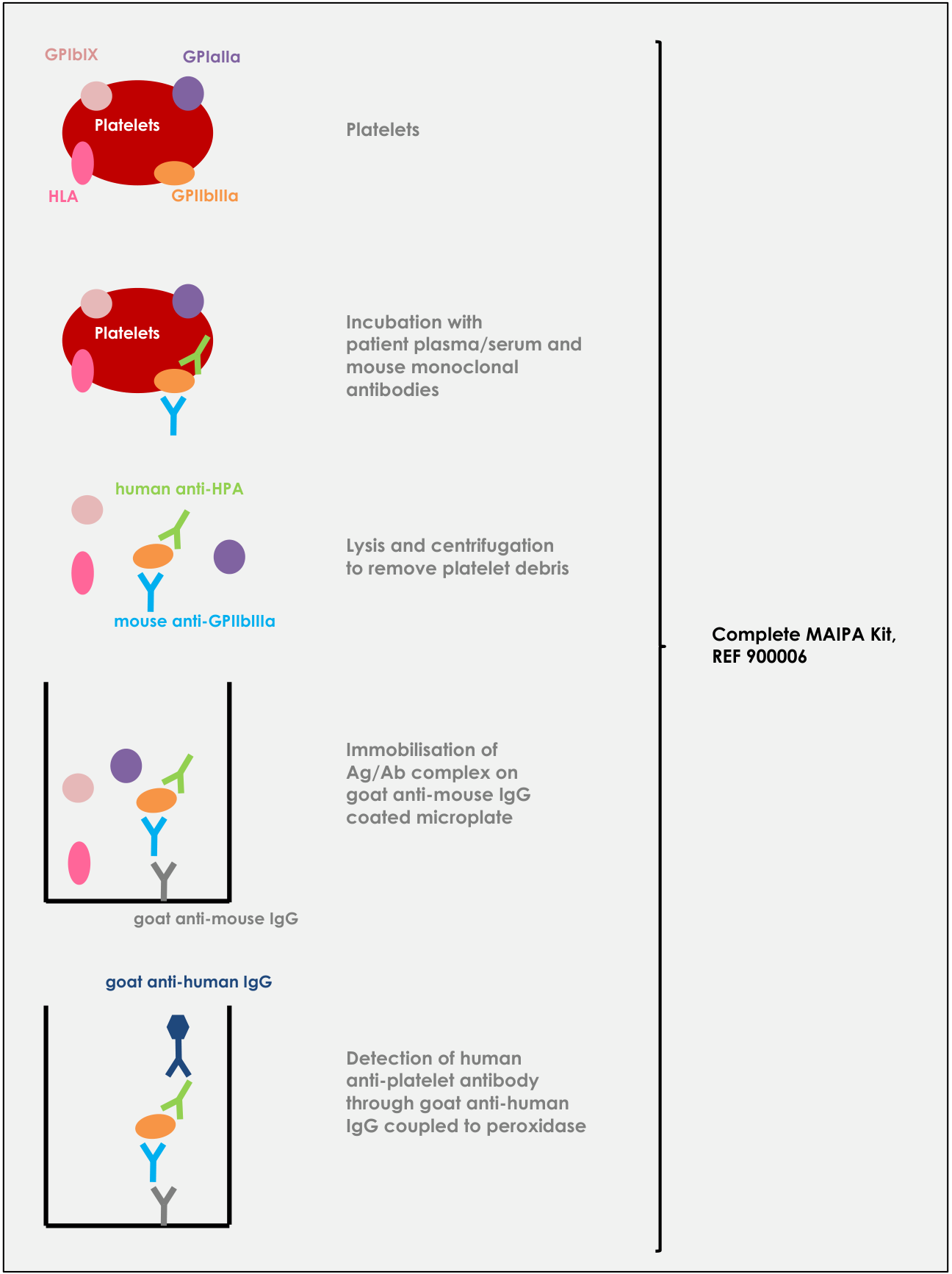
- Platelets
- Incubation with patient plasma/serum and mouse monoclonal antibodies
- Lysis and centrifugation to remove platelet debris
- Immobilisation of Ag/Ab complex on goat anti-mouse IgG coated microplate
- Detection of human anti-platelet antibody through goat anti-human IgG coupled to peroxidase
Complete MAIPA Kit, REF 900006
Concise version of the MAIPA procedure
| Indirect screening assay | Direct screening assay | Identification assay |
|---|---|---|
| 50 µL screening platelets | 50 µL patient platelets | 50 µL identification platelets |
| 3 min. 1050 g (+/- 50 g) decant shake 2 x 10 sec. 700-1000 rpm | 3 min. 1050 g (+/- 50 g) decant shake 2 x 10 sec. 700-1000 rpm | 3 min. 1050 g (+/- 50 g) decant shake 2 x 10 sec. 700-1000 rpm |
| wash 2x: 200 µL CELL WASH BUFFER 1x 3 min. 1050 g (+/- 50 g) decant shake 2 x 10 sec. 700-1000 rpm | wash 2x: 200 µL CELL WASH BUFFER 1x 3 min. 1050 g (+/- 50 g) decant shake 2 x 10 sec. 700-1000 rpm | wash 2x: 200 µL CELL WASH BUFFER 1x 3 min. 1050 g (+/- 50 g) decant shake 2 x 10 sec. 700-1000 rpm |
| 50 µL patient serum/plasma | 50 µL CELL WASH BUFFER 1x | 50 µL patient serum/plasma |
| 30 min. 36 ± 1 °C | 30 min. 36 ± 1 °C | 30 min. 36 ± 1 °C |
| 3 min. 1050 g (+/- 50 g) decant shake 2 x 10 sec. 700-1000 rpm | 3 min. 1050 g (+/- 50 g) decant shake 2 x 10 sec. 700-1000 rpm | 3 min. 1050 g (+/- 50 g) decant shake 2 x 10 sec. 700-1000 rpm |
| wash 2x: 200 µL CELL WASH BUFFER 1x 3 min. 1050 g (+/- 50 g) decant shake 2 x 10 sec. 700-1000 rpm | wash 2x: 200 µL CELL WASH BUFFER 1x 3 min. 1050 g (+/- 50 g) decant shake 2 x 10 sec. 700-1000 rpm | wash 2x: 200 µL CELL WASH BUFFER 1x 3 min. 1050 g (+/- 50 g) decant shake 2 x 10 sec. 700-1000 rpm |
| 50 µL monoclonal antibody MAB | 50 µL monoclonal antibody MAB | 50 µL monoclonal antibody MAB |
| 30 min. 36 ± 1 °C | 30 min. 36 ± 1 °C | 30 min. 36 ± 1 °C |
| 3 min. 1050 g (+/- 50 g) decant shake 2 x 10 sec. 700-1000 rpm | 3 min. 1050 g (+/- 50 g) decant shake 2 x 10 sec. 700-1000 rpm | 3 min. 1050 g (+/- 50 g) decant shake 2 x 10 sec. 700-1000 rpm |
| wash 4x: 200 µL CELL WASH BUFFER 1x 3 min. 1050 g (+/- 50 g) decant shake 2 x 10 sec. 700-1000 rpm | wash 4x: 200 µL CELL WASH BUFFER 1x 3 min. 1050 g (+/- 50 g) decant shake 2 x 10 sec. 700-1000 rpm | wash 4x: 200 µL CELL WASH BUFFER 1x 3 min. 1050 g (+/- 50 g) decant shake 2 x 10 sec. 700-1000 rpm |
| 130 µL LYSIS BUFFER mix vigorously by pipetting up and down | 130 µL LYSIS BUFFER mix vigorously by pipetting up and down | 130 µL LYSIS BUFFER mix vigorously by pipetting up and down |
| minimum 15 min. 2 - 8 °C | minimum 15 min. 2 - 8 °C | minimum 15 min. 2 - 8 °C |
| 15 min. 1050 x g (+/- 50 g) | 15 min. 1050 x g (+/- 50 g) | 15 min. 1050 x g (+/- 50 g) |
| 100 µL lysate in coated MTP | 100 µL lysate in coated MTP | 100 µL lysate in coated MTP |
| 30 min. 36 ± 1 °C | 30 min. 36 ± 1 °C | 30 min. 36 ± 1 °C |
| decant wash 6x: 200 µL ELISA WASH BUFFER 1x empty wells and tap on absorbent paper | decant wash 6x: 200 µL ELISA WASH BUFFER 1x empty wells and tap on absorbent paper | decant wash 6x: 200 µL ELISA WASH BUFFER 1x empty wells and tap on absorbent paper |
| 100 µL HRP conjugate | 100 µL HRP conjugate | 100 µL HRP conjugate |
| 30 min. 36 ± 1 °C | 30 min. 36 ± 1 °C | 30 min. 36 ± 1 °C |
| decant wash 6x: 200 µL ELISA WASH BUFFER 1x empty wells and tap on absorbent paper | decant wash 6x: 200 µL ELISA WASH BUFFER 1x empty wells and tap on absorbent paper | decant wash 6x: 200 µL ELISA WASH BUFFER 1x empty wells and tap on absorbent paper |
| 100 µL TMB substrate | 100 µL TMB substrate | 100 µL TMB substrate |
| 15 min. 36 ± 1 °C | 15 min. 36 ± 1 °C | 15 min. 36 ± 1 °C |
| 100 µL stop solution | 100 µL stop solution | 100 µL stop solution |
| read 450/620 nm | read 450/620 nm | read 450/620 nm |
Examples of MAIPA Worksheets
| Haltbarkeit / Hinweis | 1 | 2 | 3 | 4 | 5 | 6 | 7 | 8 | 9 | 10 | 11 | 12 |
|---|---|---|---|---|---|---|---|---|---|---|---|---|
| A | BLANK | PLTL PATIENT 1 no serum MAB IIbIIIa | PLTL PATIENT 2 no serum MAB IIbIIIa | PLTL PATIENT 3 no serum MAB IIbIIIa | PLTL PATIENT 4 no serum MAB IIbIIIa | PLTL PATIENT 5 no serum MAB IIbIIIa | ||||||
| B | BLANK | PLTL PATIENT 1 no serum MAB IaIIa | PLTL PATIENT 2 no serum MAB IaIIa | PLTL PATIENT 3 no serum MAB IaIIa | PLTL PATIENT 4 no serum MAB IaIIa | PLTL PATIENT 5 no serum MAB IaIIa | ||||||
| C | SCREEN PLTL CONTR 1a MAB IIbIIIa | PLTL PATIENT 1 no serum MAB HLA | PLTL PATIENT 2 no serum MAB HLA | PLTL PATIENT 3 no serum MAB HLA | PLTL PATIENT 4 no serum MAB HLA | PLTL PATIENT 5 no serum MAB HLA | ||||||
| D | SCREEN PLTL CONTR 5b MAB IaIIa | PLTL PATIENT 1 no serum MAB IbIX | PLTL PATIENT 2 no serum MAB IbIX | PLTL PATIENT 3 no serum MAB IbIX | PLTL PATIENT 4 no serum MAB IbIX | PLTL PATIENT 5 no serum MAB IbIX | ||||||
| E | SCREEN PLTL CONTR HLA MAB HLA | SCREEN PLTL SERUM PATIENT 1 MAB IIbIIIa | SCREEN PLTL SERUM PATIENT 2 MAB IIbIIIa | SCREEN PLTL SERUM PATIENT 3 MAB IIbIIIa | SCREEN PLTL SERUM PATIENT 4 MAB IIbIIIa | SCREEN PLTL SERUM PATIENT 5 MAB IIbIIIa | ||||||
| F | SCREEN PLTL CONTR NEG MAB IbIX | SCREEN PLTL SERUM PATIENT 1 MAB IaIIa | SCREEN PLTL SERUM PATIENT 2 MAB IaIIa | SCREEN PLTL SERUM PATIENT 3 MAB IaIIa | SCREEN PLTL SERUM PATIENT 4 MAB IaIIa | SCREEN PLTL SERUM PATIENT 5 MAB IaIIa | ||||||
| G | SCREEN PLTL SERUM PATIENT 1 MAB HLA | SCREEN PLTL SERUM PATIENT 2 MAB HLA | SCREEN PLTL SERUM PATIENT 3 MAB HLA | SCREEN PLTL SERUM PATIENT 4 MAB HLA | SCREEN PLTL SERUM PATIENT 5 MAB HLA | |||||||
| H | SCREEN PLTL SERUM PATIENT 1 MAB IbIX | SCREEN PLTL SERUM PATIENT 2 MAB IbIX | SCREEN PLTL SERUM PATIENT 3 MAB IbIX | SCREEN PLTL SERUM PATIENT 4 MAB IbIX | SCREEN PLTL SERUM PATIENT 5 MAB IbIX |
| Haltbarkeit / Hinweis | 1 | 2 | 3 | 4 | 5 | 6 | 7 | 8 | 9 | 10 | 11 | 12 |
|---|---|---|---|---|---|---|---|---|---|---|---|---|
| A | BLANK | ID PLTL 1 SERUM PATIENT 1 MAB IIbIIIa | ID PLTL 1 SERUM PATIENT 2 MAB IIbIIIa | ID PLTL 1 SERUM PATIENT 3 MAB IIbIIIa | ID PLTL 1 SERUM PATIENT 4 MAB IIbIIIa | ID PLTL 1 SERUM PATIENT 5 MAB IaIIa | ||||||
| B | BLANK | ID PLTL 2 SERUM PATIENT 1 MAB IIbIIIa | ID PLTL 2 SERUM PATIENT 2 MAB IIbIIIa | ID PLTL 2 SERUM PATIENT 3 MAB IIbIIIa | ID PLTL 2 SERUM PATIENT 4 MAB IIbIIIa | ID PLTL 2 SERUM PATIENT 5 MAB IaIIa | ||||||
| C | SCREEN PLTL CONTR 1a MAB IIbIIIa | ID PLTL 3 SERUM PATIENT 1 MAB IIbIIIa | ID PLTL 3 SERUM PATIENT 2 MAB IIbIIIa | ID PLTL 3 SERUM PATIENT 3 MAB IIbIIIa | ID PLTL 3 SERUM PATIENT 4 MAB IIbIIIa | ID PLTL 3 SERUM PATIENT 5 MAB IaIIa | ||||||
| D | SCREEN PLTL CONTR 5b MAB IaIIa | ID PLTL 4 SERUM PATIENT 1 MAB IIbIIIa | ID PLTL 4 SERUM PATIENT 2 MAB IIbIIIa | ID PLTL 4 SERUM PATIENT 3 MAB IIbIIIa | ID PLTL 4 SERUM PATIENT 4 MAB IIbIIIa | ID PLTL 4 SERUM PATIENT 5 MAB IaIIa | ||||||
| E | SCREEN PLTL CONTR HLA MAB HLA | ID PLTL 5 SERUM PATIENT 1 MAB IIbIIIa | ID PLTL 5 SERUM PATIENT 2 MAB IIbIIIa | ID PLTL 5 SERUM PATIENT 3 MAB IIbIIIa | ID PLTL 5 SERUM PATIENT 4 MAB IIbIIIa | ID PLTL 5 SERUM PATIENT 5 MAB IaIIa | ||||||
| F | SCREEN PLTL CONTR NEG MAB IbIX | ID PLTL 6 SERUM PATIENT 1 MAB IIbIIIa | ID PLTL 6 SERUM PATIENT 2 MAB IIbIIIa | ID PLTL 6 SERUM PATIENT 3 MAB IIbIIIa | ID PLTL 6 SERUM PATIENT 4 MAB IIbIIIa | ID PLTL 6 SERUM PATIENT 5 MAB IaIIa | ||||||
| G | ID PLTL 1 SERUM PATIENT 6 MAB IaIIa | ID PLTL 2 SERUM PATIENT 6 MAB IaIIa | ID PLTL 3 SERUM PATIENT 6 MAB IaIIa | ID PLTL 4 SERUM PATIENT 6 MAB IaIIa | ID PLTL 5 SERUM PATIENT 6 MAB IaIIa | ID PLTL 6 SERUM PATIENT 6 MAB IaIIa | ||||||
| H | ID PLTL 1 SERUM PATIENT 7 MAB IaIIa | ID PLTL 2 SERUM PATIENT 7 MAB IaIIa | ID PLTL 3 SERUM PATIENT 7 MAB IaIIa | ID PLTL 4 SERUM PATIENT 7 MAB IaIIa | ID PLTL 5 SERUM PATIENT 7 MAB IaIIa | ID PLTL 6 SERUM PATIENT 7 MAB IaIIa |
| Haltbarkeit / Hinweis | 1 | 2 | 3 | 4 | 5 | 6 | 7 | 8 | 9 | 10 | 11 | 12 |
|---|---|---|---|---|---|---|---|---|---|---|---|---|
| A | BLANK | ID PLTL 1 SERUM PATIENT 1 MAB IIbIIIa | ID PLTL 1 SERUM PATIENT 2 MAB IIbIIIa | PLTL PATIENT 4 no serum MAB IIbIIIa | PLTL PATIENT 5 no serum MAB IIbIIIa | PLTL PATIENT 6 no serum MAB IIbIIIa | ||||||
| B | BLANK | ID PLTL 2 SERUM PATIENT 1 MAB IIbIIIa | ID PLTL 2 SERUM PATIENT 2 MAB IIbIIIa | PLTL PATIENT 4 no serum MAB IaIIa | PLTL PATIENT 5 no serum MAB IaIIa | PLTL PATIENT 6 no serum MAB IaIIa | ||||||
| C | SCREEN PLTL CONTR 1a MAB IIbIIIa | ID PLTL 3 SERUM PATIENT 1 MAB IIbIIIa | ID PLTL 3 SERUM PATIENT 2 MAB IIbIIIa | PLTL PATIENT 4 no serum MAB HLA | PLTL PATIENT 5 no serum MAB HLA | PLTL PATIENT 6 no serum MAB HLA | ||||||
| D | SCREEN PLTL CONTR 5b MAB IaIIa | ID PLTL 4 SERUM PATIENT 1 MAB IIbIIIa | ID PLTL 4 SERUM PATIENT 2 MAB IIbIIIa | PLTL PATIENT 4 no serum MAB IbIX | PLTL PATIENT 5 no serum MAB IbIX | PLTL PATIENT 6 no serum MAB IbIX | ||||||
| E | SCREEN PLTL CONTR HLA MAB HLA | ID PLTL 5 SERUM PATIENT 1 MAB IIbIIIa | ID PLTL 5 SERUM PATIENT 2 MAB IIbIIIa | SCREEN PLTL SERUM PATIENT 4 MAB IIbIIIa | SCREEN PLTL SERUM PATIENT 5 MAB IIbIIIa | SCREEN PLTL SERUM PATIENT 6 MAB IIbIIIa | ||||||
| F | SCREEN PLTL CONTR NEG MAB IbIX | ID PLTL 6 SERUM PATIENT 1 MAB IIbIIIa | ID PLTL 6 SERUM PATIENT 2 MAB IIbIIIa | SCREEN PLTL SERUM PATIENT 4 MAB IaIIa | SCREEN PLTL SERUM PATIENT 5 MAB IaIIa | SCREEN PLTL SERUM PATIENT 6 MAB IaIIa | ||||||
| G | ID PLTL 1 SERUM PATIENT 3 MAB IaIIa | ID PLTL 3 SERUM PATIENT 3 MAB IaIIa | ID PLTL 5 SERUM PATIENT 3 MAB IaIIa | SCREEN PLTL SERUM PATIENT 4 MAB HLA | SCREEN PLTL SERUM PATIENT 5 MAB HLA | SCREEN PLTL SERUM PATIENT 6 MAB HLA | ||||||
| H | ID PLTL 2 SERUM PATIENT 3 MAB IaIIa | ID PLTL 4 SERUM PATIENT 3 MAB IaIIa | ID PLTL 6 SERUM PATIENT 3 MAB IaIIa | SCREEN PLTL SERUM PATIENT 4 MAB IbIX | SCREEN PLTL SERUM PATIENT 5 MAB IbIX | SCREEN PLTL SERUM PATIENT 6 MAB IbIX |
MAIPA Worksheet Template
| Haltbarkeit / Hinweis | 1 | 2 | 3 | 4 | 5 | 6 | 7 | 8 | 9 | 10 | 11 | 12 |
|---|---|---|---|---|---|---|---|---|---|---|---|---|
| A | BLANK | |||||||||||
| B | BLANK | |||||||||||
| C | SCREEN PLTL CONTR 1a MAB IIbIIIa | |||||||||||
| D | SCREEN PLTL CONTR 5b MAB IaIIa | |||||||||||
| E | SCREEN PLTL CONTR HLA MAB HLA | |||||||||||
| F | SCREEN PLTL CONTR NEG MAB IbIX | |||||||||||
| G | ||||||||||||
| H |
Revision History
| Version number | Description |
|---|---|
| IFU900006/V17/11-2021 | Additions to ‘Warnings and precautions’ section (clauses 4.2, 4.4, 4.5, 4.6, 4.7, 4.10, 4.11). Additions on required equipment, specifically on centrifuges in section 8. Restructure of MAIPA procedure description: • Division in separate instructions for Indirect and Direct MAIPA • Inclusion of a concise version of the complete MAIPA procedure in Annex. Application of the legal company name of the manufacturer: ‘Advanced Practical Diagnostics BV’ in stead of the abbreviation ‘apDia’. |
| IFU900006/V18/02-2023 | Addition to sections 9 and 10 « Test Procedure Indirect/Direct MAIPA » (clauses 9.5 and 10.4) : for validation of a MAIPA run, the negative control of the kit may not only be tested with detection antibody anti-GPIbIX but antibodies anti-GPIIbIIIa or anti-GPIaIIa can be used as well. |
| IFU900006/V19/07-2023 | Changes related to the implementation of the IVDR requirements for Class C devices: • Identification number of notified body affixed to CE mark pictogram • Adding “Intended purpose” header + rephrasing “Definition and scope” • In section 1 “Reagents”: omission of the antibody concentration (10 µl) of the detection antibodies; antibody concentration can be batch dependent. • In section 4 “Warnings and precautions for users”: addition of applicable hazard pictograms. • In section 5 “Sample Material”: addition of statement related to allowed number of freeze-thaw cycles for frozen samples • Extended description of the test characteristics in section on ”Performance Characteristics” of the MAIPA Assay • Inclusion of a new section related to “Summary of Safety and Performance” • In “Bibliography”: addition of a reference to chloroquine treatment |