















Produktinformationen
Faktor IX
Aus dem Original-PDF übertragen und mit ihm abgeglichen. Maßgeblich bleibt das Originaldokument.
For In Vitro Diagnostic Use – 2 x 50 tests
ENGLISH – Insert Revision 04/2014
INTENDED USE
For determination of Factor IX (FIX) activity in plasma and FIX containing concentrates.
BIOCHEMISTRY
FIX is a single chain, vitamin K dependent glycoprotein of about 55 KDa, which is activated by FXIa or tissue factor / FVIIa. Activated FIX (FIXa) converts FX to FXa in the presence of FVIII, phospholipids and calcium ions.
MEASUREMENT PRINCIPLE
FIX activity is determined in a chromogenic method, in which human FIX is activated by human FXIa and where formed FIXa activates human FX in the presence of human FVIII, calcium ions and phospholipid. Similar to in vivo conditions, FVIII is activated by thrombin which is generated during the incubation. The amount of FXa formed is related to the FIX activity and is determined from the hydrolysis of a chromogenic FXa substrate. The FIX activity of the sample is assigned vs. a FIX plasma or a FIX concentrate standard with FIX potency expressed in International Units (IU).
KIT COMPOSITION
Reagent A (2 vials) – REF 9010
Reagent A contains lyophilized human FVIII, human FX, bovine FV and a fibrin polymerization inhibitor.
Reagent B (2 vials) – REF 9020
Reagent B contains lyophilized human FXIa, human FII, calcium chloride and phospholipids.
FXa Substrate, 6 mL (1 vial) – REF 9080
Liquid solution of chromogenic FXa substrate (Z-D-Arg-Gly-Arg-pNA), 2.5 mmol/L, containing a thrombin inhibitor.
FIX Diluent Buffer, Stock Solution, 20 mL (1 vial) – REF 9050
Liquid stock solution of diluent buffer, containing a heparin antagonist.
PRECAUTIONS AND WARNINGS
The reagents are matched – only use reagents from the same kit lot.
CAUTION: Each donor unit used in the reagents has been tested by FDA approved methods for the presence of Hepatitis B surface antigen and antibodies to HIV 1 and 2 and Hepatitis C and found to be negative. However, since no test can completely rule out the presence of these blood borne diseases, the handling and disposal of these human sourced reagents should be handled with the required caution, as being potentially infectious.
PREPARATION
Reagent A
Reconstitute with 1.4 mL water. Allow to stand for 5 min at 20-25°C with intermittent gentle mixing for complete reconstitution.
Reagent B
Reconstitute with 8.0 mL water. Allow to stand for 5 min at 20-25°C with intermittent gentle mixing for complete reconstitution.
FXa Substrate, 6 mL
Ready for use.
FIX Diluent Buffer, Stock Solution, 20 mL
Dilute 1 + 9 with water to obtain a 0.025 mol/L Tris-HCl buffer working solution, pH 7.9 (at 20°C), with 1% bovine serum albumin and a heparin antagonist. NB: The vial is slightly over dispensed. Always measure up the desired volume prior to 10-fold dilution with water.
Note: All reconstitutions and dilutions should be made with water of a quality of at least NCCLS Type II water or Ph Eur water for injection.
STORAGE AND STABILITY
The sealed reagents are stable at 2-8°C until the Expiry Date printed on the label. Be careful to avoid contamination of the reagents by microorganisms.
- Reagent A: Stability after reconstitution is 72 hours at 2-8°C, 8 h at 20-25°C and 12 months at ≤ -70°C.
- Reagent B: Stability after reconstitution is 72 hours at 2-8°C, 8 h at 37°C and 12 months at ≤ -70°C.
- Chromogenic FXa substrate: Opened vial is stable for 1 month at 2-8°C and 12 months at ≤ -20°C.
- FIX Diluent Buffer Stock Solution: Opened vial is stable for 1 month at 2-8°C. Buffer working solution should be used the same day as prepared.
MATERIALS REQUIRED BUT NOT PROVIDED
- Deionized water, NCCLS Type II water or Ph. Eur. Water for injection or higher quality
- For calibration: Human plasma or FIX concentrate, potency assigned vs. a WHO International Standard for FIX activity
- Citric acid, 2% (for end-point method)
- Calibrated pipettes
- Photometer, 405 nm (and 490 nm for end-point method)
- Heat incubator 37°C
- Plastic test tubes
- Stop-watch
- Vortex mixer
For microplate assay, make sure to use low binding microplates.
SYMBOLS USED
| Symbol | Meaning |
|---|---|
| IVD | In Vitro Diagnostic Medical Device |
| REF | Catalogue number |
| LOT | Batch code |
| Hourglass | Use by |
| Thermometer | Temperature limitation |
| Book with “i” | Consult instruction for use |
| Biohazard | Biological risks |
| Factory | Manufacturer |
| CE | CE Mark |
SPECIMEN COLLECTION AND TREATMENT
Sample collection must be in conformity with the recommendations for haemostasis tests. Freshly drawn venous blood (9 volumes) is collected into 0.109 M trisodium citrate anticoagulant (1 volume). Use silicon glass or a plastic test tube. Centrifuge for 15 min at 2000-2500 g. Refer to CLSI guideline H21-A5 for further instruction on specimen collection, handling and storage.
QUALITY CONTROL
Quality control plasmas with assigned FIX activity are commercially available and should be used for validating the calibration curve. Normal and abnormal controls are recommended for a complete quality control program. The controls should be processed as the samples. Each laboratory should determine its own quality control range, either by means of the target values and ranges provided by the manufacturer of the controls or by means of its own confidence level established in the laboratory.
METHOD - PLASMA
A calibration curve should be included in each run. A normal human plasma calibrated against an International Standard should be used as calibrator.
Prepare standard dilutions in FIX Diluent Buffer working solution to obtain standards in the selected range. Prepare at least five different standard dilutions. It is recommended to prepare independent dilutions of each standard.
All dilutions should be prepared in plastic test tubes.
12.1 High range (25 - 200 %) with sample dilution 1:80
Example - Standard dilutions, high range:
| FIX Standard % | Total Dilution | Volume | Volume of FIX Diluent Buffer working solution |
|---|---|---|---|
| Predilution | 1:10 | 100 µL of plasma | 900 µL |
| 200% | 1:40 | 100 µL of predilution | 300 µL |
| 150% | 1:53.3 | 100 µL of predilution | 433 µL |
| 100% | 1:80 | 100 µL of predilution | 700 µL |
| 50% | 1:160 | 50 µL of predilution | 750 µL |
| 25% | 1:320 | 50 µL of predilution | 1550 µL |
| Reagent Blank | - | - | 500 µL |
NOTE: 100% activity is defined as a FIX activity of 1 IU/mL in plasma. In case the FIX activity of the plasma standard differs from this value, an appropriate correction factor should be used when calculating the sample result. It is recommended to express all sample results as IU/mL.
12.2 Sample dilution – High range
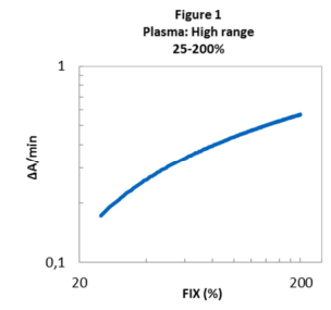
Plasma samples with an estimated potency of 25 - 200 % (0.25 - 2 IU/mL) should be analysed in the high range, using sample dilution 1:80. The FIX activity of the tested sample is obtained directly from the calibration curve.
12.3 Low range (0.5 - 25 %) with sample dilution 1:20
Example – Standard dilutions, low range:
| FIX Standard % | Total Dilution | Volume | Volume of FIX Diluent Buffer working solution |
|---|---|---|---|
| Predilution | 1:20 | 50 µL of plasma | 950 µL |
| 25% | 1:80 | 100 µL of predilution | 300 µL |
| 12.5% | 1:160 | 100 µL of predilution | 700 µL |
| 6.25% | 1:320 | 100 µL of predilution | 1500 µL |
| 3.125% | 1:640 | 50 µL of predilution | 1550 µL |
| 1 % | 1:2000 | 10 µL of predilution | 990 µL |
| 0.5 % | 1:4000 | 10 µL of predilution | 1990 µL |
| Reagent Blank | - | - | 500 µL |
NOTE: 100% activity is defined as a FIX activity of 1 IU/mL in plasma. In case the FIX activity of the plasma standard differs from this value, an appropriate correction factor should be used when calculating the sample result. It is recommended to express all sample results as IU/mL.
12.3.1 Sample dilution - Low range
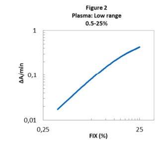
Plasma samples with an estimated potency of 0.5 - 25% (0.005 - 0.25 IU/mL) should be analysed in the low range, using sample dilution 1:20. The FIX activity of the tested sample is obtained directly from the calibration curve.
12.3.2 Sample Blank (End-point method only)
Sample blanks are in general not necessary but should be included when analyzing hemolytic, icteric or lipemic plasma samples using the end-point method. A sample blank is obtained by adding Citric Acid prior to the other reagents.
METHOD – CONCENTRATES
The European Pharmacopoeia recommends parallel line analysis for potency assignment of biological samples. FIX concentrates could be analyzed in the range 0.25 - 200 mIU/mL using a 4- or 5-parameter curve fit or in the range 0.5 – 5 mIU/mL using log-log transformation and a linear curve fit. For parallel line analysis it is recommended that each laboratory establishes its own range for potency assignments.
13.1 Standard dilutions – FIX containing concentrates
A calibration curve should be included in each run. An international reference material for FIX concentrates or an internal or commercially available FIX concentrate, calibrated against an international standard, should be used as calibrator.
Prepare standard dilutions in FIX Diluent Buffer working solution to obtain standards within the range 0.25 - 200 mIU/mL. Prepare at least five different standard dilutions. All dilutions should be prepared in plastic test tubes.
13.2 Sample dilution - FIX containing concentrates
It is recommended to prepare at least three sample dilutions in FIX Diluent Buffer working solution with activities within the standard range. All dilutions should be prepared in plastic test tubes.
ASSAY PROTOCOL– PLASMA AND CONCENTRATES
14.1 Manual method
The same assay procedure should be used for both plasma high range and low range as well as for concentrates.
| Step | Volume |
|---|---|
| Sample / Standard dilution | 25 µL |
| Reagent A | 25 µL |
| Incubation 3-4 min, 37°C | |
| Reagent B (37°C) | 150 µL |
| Activation 8 min, 37°C | |
| FXa Substrate (37°C) | 50 µL |
| Kinetic method: Read ΔA405/min at 37°C End-point method: Hydrolysis at 37°C for 2 min | |
| Citric Acid, 2% (End-point method only) | 50 µL |
Kinetic reading:
Read the absorbance at 405 nm and record the change in absorbance.
End-point method:
Stop the reaction with 2% citric acid. Read the absorbance at 405 nm, using 490 nm as reference wavelength. Absorbance readings should be made within 2 hours after termination of the substrate hydrolysis.
14.2 Automated methods
Protocols for various automated coagulation instruments are available upon request.
Special note, Reagent B: For stability reasons, only use siliconized glass vials or plastic vials if a reagent vial needs to be replaced in order to fit the analyzer.
CALCULATION
Plasmas:
- Plot the maximal absorbance change/minute (ΔA405ₘₐₓ/min) or absorbance (A405-490) vs. FIX activity in a Log-Log graph after subtracting the reagent blank (Fig 1 and Fig 2). Alternatively, a Semi-Log graph with a 4- or 5-parameter curve fit may be used.
- The FIX activity of the tested sample is obtained directly from the calibration curve. Correct the obtained value with the appropriate correction factor if the FIX activity of the normal plasma standard differs from 1 IU/mL.
- Adjust for the dilution factor if several dilutions are used.
- Express the sample result as IU/mL or %.
Concentrates:
- Plot the maximal absorbance change/minute (ΔA405ₘₐₓ/min) or absorbance (A405-490) vs. FIX activity in a Semi-Log or Log-Log graph after subtracting the reagent blank. Use a 4 or 5-parameter curve fit (Semi-Log) or linear curve fit (Log-Log.
- Determine the FIX activity of the sample from the calibration curve using the parallel line model.
- Adjust for the dilution used and express the sample results as IU/mL.
- The European Pharmacopoeia recommends use of the parallel line model. Alternatively, the Factor IX activity in each dilution of the tested sample can be directly obtained from the calibration curve. The result should then be multiplied by the dilution used.
EXPECTED VALUES
The Factor IX levels measured in 25 healthy males and 25 healthy females, aged between 19 and 56 were in the range 0.6 - 1.3 IU/mL. FIX Deficiency, also known as Haemophilia B, can be divided into three categories³: Mild (0.05 - 0.4 IU/mL), moderate (0.01 - 0.05 IU/mL) and severe (≤0.01 IU/mL). FIX levels can be decreased in patients with hepatic disease, cirrhosis and DIC as well as in patients on anti-vitamin K therapy.
PERFORMANCE CHARACTERISTICS
Detection limit: 0.1% (0.001 IU/mL), calculated according to CLSI EP17-A using the low range and sample dilution of 1:20.
Quantification limit: 0.5% (0.005 IU/mL), calculated according to CLSI EP17-A using the low range and sample dilution 1:20.
Precision:
Repeatability (Intra assay CV): 3% Within Laboratory (Inter assay CV): 4% The precision was determined at 1%, 25% and 100% Factor IX activity. The results were obtained using a manual microplate method.
Linearity: 0.5 – 200% (0.005 – 2 IU/mL), calculated according to CLSI EP06-A.
INTERFERENCE
FIX results are not affected by plasma concentrations up to: Hemoglobin - 5 g/L, Bilirubin - 0.4 g/L, Triglycerides - 5 g/L, LMW Heparin - 5 IU/mL, and unfractionated Heparin - 2 U/mL. There is no interference of FIXa up to 50 mIU FIXa/1 IU FIX.
REFERENCES
- Gray E, Barson H, Hockley J, Rigsby P. Collaborative study for value assignment of the 4ᵗʰ International Standard for Factors II, VII, IX, X, Plasma. WHO/BS/10.2145; 2010: 1-43.
- Pickering WM, Gray E. The effect of activated Factor IX on the Factor IX coagulant and NAPTT activity of high-purity Factor IX concentrates. J. Thromb Haemost 2007; 5, Supplement 2: P-T-156.
- White GC et al. Definitions in Hemophilia. Thromb Haemost 2001; 85: 560.
- Bertina RM. Elevated clotting factor levels and venous thrombosis. Pathophysiol Haemost Thromb 2003/2004; 33: 395-400.
- Clinical and Laboratory Standards Institute (CLSI), www.clsi.org
- 6ᵗʰ Edition of the European Pharmacopoeia, General Chapter 5.3 Statistical analysis of results of biological assays and tests.
Manufacturer
Rossix AB Taljegårdsgatan 3B SE-431 53 Mölndal, Sweden info@rossix.com www.rossix.com